
 400-115-0588
 在线咨询
在线咨询- 029-86354885
- 18392009562
| Catalog # | Pkg Size | Price | Quantity | Shopping Cart |
|---|---|---|---|---|
| R-POSN1604 | 1mg | ¥ 1950 | 加入购物车 | |
| R-POSN1604 | 5mg | ¥ 7800 | 加入购物车 | |
 Datasheet
 MSDS
MSDS COA
COA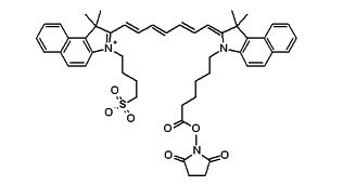
| 外观 | 棕红色粉末 |
| 分子量 | N/A |
| 溶解度 | DMSO |
| CAS号 | N/A |
| 质量控制 | >90% |
| 储存条件 | -20℃ |
| 保存时间 | 1年 |
| 其他信息 | Ex/Em(nm):780/800 |
1. Prepare protein stock solution (Solution A):
Mix 100 µL of a reaction buffer (e.g., 1 M sodium carbonate solution or 1 M phosphate buffer with pH ~9.0) with 900 µL of the target protein solution (e.g. antibody, protein concentration >2 mg/ml if possible) to give 1 mL protein labeling stock solution.
Note 1: The pH of the protein solution (Solution A) should be 8.5 ± 0.5. If the pH of the protein solution is lower than 8.0, adjust the pH to the range of 8.0-9.0 using 1 M sodium bicarbonate solution or 1 M pH 9.0 phosphate buffer.
Note 2: The protein should be dissolved in 1X phosphate buffered saline (PBS), pH 7.2-7.4. If the protein is dissolved in Tris or glycine buffer, it must be dialyzed against 1X PBS, pH 7.2-7.4, to remove free amines or ammonium salts (such as ammonium sulfate and ammonium acetate) that are widely used for protein precipitation.
Note 3: Impure antibodies or antibodies stabilized with bovine serum albumin (BSA) or gelatin will not be labeled well. The presence of sodium azide or thimerosal might also interfere with the conjugation reaction. Sodium azide or thimerosal can be removed by dialysis or spin column for optimal labeling results.
Note 4: The conjugation efficiency is significantly reduced if the protein concentration is less than 2 mg/mL. For optimal labeling efficiency the final protein concentration range of 2-10 mg/mL is recommended.
2. Prepare dye stock solution (Solution B):
Add anhydrous DMSO into the vial of ICG dyes to make a 10-20 mM stock solution. Mix well by pipetting or vortex.
Note: Prepare the dye stock solution (Solution B) before starting the conjugation. Use promptly. Extended storage of the dye stock solution may reduce the dye activity. Solution B can be stored in freezer for two weeks when kept from light and moisture. Avoid freeze-thaw cycles.
3. Determine the optimal dye/protein ratio (optional):
Note: Each protein requires distinct dye/protein ratio, which also depends on the properties of dyes. Over labeling of a protein could detrimentally affects its binding affinity while the protein conjugates of low dye/protein ratio gives reduced sensitivity. We recommend you experimentally determine the best dye/protein ratio by repeating Steps 4 and 5 using a serial different amount of labeling dye solutions. In general 4-6 dyes/protein are recommended for most of dye-protein conjugates.
3.1 Use 10:1 molar ratio of Solution B (dye)/Solution A (protein) as the starting point: Add 5 µl of the dye stock solution (Solution B, assuming the dye stock solution is 10 mM) into the vial of the protein solution (95 µl of Solution A) with effective shaking. The concentration of the protein is ~0.05 mM assuming the protein concentration is 10 mg/mL and the molecular weight of the protein is ~200KD.
Note: The concentration of the DMSO in the protein solution should be <10 %.
3.2 Run conjugation reaction (see Step 4 below).
3.3 Repeat #3.2 with the molar ratios of Solution B/Solution A at 5:1; 15:1 and 20:1 respectively.
3.4 Purify the desired conjugates using premade spin columns.
3.5 Calculate the dye/protein ratio (DOS) for the above 4 conjugates (see next page).
3.6 Run your functional tests of the above 4 conjugates to determine the best dye/protein ratio to scale up your labeling reaction.
4. Run conjugation reaction:
4.1 Add the appropriate amount of dye stock solution (Solution B) into the vial of the protein solution (Solution A) with effective shaking.
Note: The best molar ratio of Solution B/Solution is determined from Step 3.6. If Step 3 is skipped, we recommend using 10:1 molar ratio of Solution B (dye)/Solution A (protein).
4.2 Continue to rotate or shake the reaction mixture at room temperature for 30-60 minutes.
5. Purify the conjugation
The following protocol is an example of dye-protein conjugate purification by using a Sephadex G-25 column.
5.1 Prepare Sephadex G-25 column according to the manufacture instruction.
5.2 Load the reaction mixture (directly from Step 4) to the top of the Sephadex G-25 column.
5.3 Add PBS (pH 7.2-7.4) as soon as the sample runs just below the top resin surface.
5.4 Add more PBS (pH 7.2-7.4) to the desired sample to complete the column purification. Combine the fractions that contain the desired dye-protein conjugate.
Note 1: For immediate use, the dye-protein conjugate need be diluted with staining buffer, and aliquoted for multiple uses.
Note 2: For longer term storage, dye-protein conjugate solution need be concentrated or freeze dried (see below).
Characterize the Desired Dye-Protein Conjugate
The Degree of Substitution (DOS) is the most important factor for characterizing dye-labeled protein. Proteins of lower DOS usually have weaker fluorescence intensity, but proteins of higher DOS (e.g. DOS > 6) tend to have reduced fluorescence too. The optimal DOS for most antibodies is recommended between 2 and 10 depending on the properties of dye and protein. For effective labeling, the degree of substitution should be controlled to have 4-10 moles of ICG to one mole of antibody. The following steps are used to determine the DOS of ICG labeled proteins.
1. Measure absorption:
To measure the absorption spectrum of a dye-protein conjugate, it is recommended to keep the sample concentration in the range of 1-10 µM depending on the extinction coefficient of the dye.
2. Read OD (absorbance) at 280 nm and dye maximum absorption (ƛmax = 785 nm for ICG dyes):
For most spectrophotometers, the sample (from the column fractions) need be diluted with de-ionized water so that the OD values are in the range of 0.1 to 0.9. The O.D. (absorbance) at 280 nm is the maximum absorption of protein while 785 nm is the maximum absorption of ICG dyes. To obtain accurate DOS, make sure that the conjugate is free of the non-conjugated dye.
3. Calculate DOS using the following equations:
3.1 Calculate protein concentration![]()
3.2 Calculate ![]()
3.3 Calculate the degree of labeling DOS = [Dye]/[Protein] =[DOD785´Pε280] /[230000 ×(A280-0.073A785)]
[Dye] is the dye concentration, and can be readily calculated from the Bee-Lambert Law: A=εdyeCL. [Protein] is the protein concentration. This value can be either estimated by the weight (added to the reaction) if the conjugation efficiency is high enough (preferably > 70%) or more accurately calculated by the Beer-Lambert Law: A=εproteinCL. For example, IgG has the ε value to be 203,000 cm-1M-1. Pε280 ![]() protein molar extinction coefficient at 280 nm (e. g. the molar extinction coefficient of IgG is 203,000
protein molar extinction coefficient at 280 nm (e. g. the molar extinction coefficient of IgG is 203,000
cm-1M-1). CF (dye absorption correction factor at 280 nm ) = OD280/OD750 = 0.073 for ICG-Sulfo-OSu. 230,000 cm-1M-1 is the molar extinction coefficient of ICG-Sulfo-OSu.
| Cy7.5 maleimide | Cyanine7.5 maleimide is a thiol reactive near infrared dye. |
| Cy7 (CAS 943298-08-6) | 花氰染料cy7,常被应用于生物分子标记,荧光成像及其他荧光生物分析。 |
| Cy5,(CAS:146368-11-8) | 花氰染料cy5,常被应用于生物分子标记,荧光成像及其他荧光生物分析。 |
| Cy3,(CAS:146368-13-0 ) | 花氰染料cy3,常被应用于生物分子标记,荧光成像及其他荧光生物分析。 |
| Cyanine3 NHS ester | Cyanine3 NHS ester, CAS:1393363-07-9,Cy3 NHS ester可以和带有NH2氨基的多肽、蛋白、抗体、核酸或者其他小分子连接 |
| Cyanine5 NHS ester | Cyanine5 NHS ester, CAS 1032678-42-4 Cy5 NHS ester可以和带有NH2氨基的多肽、蛋白、抗体、核酸或者其他小分子连接 |


 库存查询
库存查询